Vanderbilt Health researchers receive ‘Mars Shot’ award to improve invasive breast cancer diagnosis

Submitted by vicc_news on

Vanderbilt Health researchers Adam Rosenberg, PhD, and Laura Kennedy, MD, PhD, have received a 2026 “Mars Shot” Research Award to support studies aimed at improving the diagnosis of invasive lobular carcinoma, an invasive breast cancer that is difficult to detect.

Adam Rosenberg, PhD

The $100,000 award was announced Oct. 15 by the Society of Nuclear Medicine and Molecular Imaging (SNMMI) and the Lobular Breast Cancer Alliance Inc. (LBCA), which jointly funded the award.

Approximately 48,000 women are diagnosed with invasive lobular carcinoma in the United States each year, yet the disease remains “underappreciated and under-researched,” SNMMI President-Elect Gary Ulaner, MD, PhD, said in a news release.

Rosenberg and Kennedy “will use novel molecular imaging to help open new therapeutic options to patients …, which is very good news indeed,” said Ulaner, Chair of Molecular Imaging and Therapy at the Hoag Family Cancer Institute and Professor of Radiology and Translational Genomics at the University of Southern California.

Rosenberg is Research Associate Professor of Radiology and Radiological Sciences and Director of Radiochemistry Research at the Vanderbilt University Institute of Imaging Science. Kennedy is Assistant Professor of Medicine in the Division of Hematology and Oncology. Both are members of the Vanderbilt-Ingram Cancer Center.

Laura Kennedy, MD, PhD

The focus of their research is HER2 (human epidermal growth factor receptor 2) overexpression, which in breast cancer is associated with poor outcomes. Targeted anti-HER2 antibody therapies have improved survival considerably. However, it is difficult to accurately determine the HER2 status of invasive lobular carcinoma that has spread, or metastasized.

As a result, the burden of disease is frequently underestimated; invasive lobular carcinoma is often diagnosed in later stages and with larger tumors; and many patients are excluded from clinical trials of targeted HER2 therapies such as combined trastuzumab and deruxtecan due to insufficiently measurable disease.

One promising approach is to use positron emission tomography (PET), a noninvasive imaging technique, to detect ABY-025, an anti-HER2 antibody made by the Swedish biopharmaceutical firm Affibody, which has been labeled with gallium-68, a positron-emitting radioisotope of gallium.

Rosenberg and Kennedy will use this approach to quantify HER2 expression throughout the body including bone, the lining of the abdominal cavity, and gastrointestinal sites that cannot be sampled, visualized on conventional imaging, or accurately measured by biopsy.

If this imaging technique finds “actionable HER2” in tumors that were labeled “HER2 zero” by conventional immunohistochemistry testing, “it would show molecular imaging working as a quantitative predictive biomarker that decides drug eligibility, not just as a way to locate disease,” Rosenberg said.

“At Vanderbilt-Ingram Cancer Center, we are committed to advancing precision oncology through innovative research that improves the lives of patients,” said Vanderbilt-Ingram Director Ben Ho Park, MD, PhD, the Benjamin F. Byrd Jr. Professor of Oncology and Professor of Medicine.

“Drs. Rosenberg and Kennedy are leading groundbreaking work to better understand HER2 expression in invasive lobular carcinoma, with the potential to expand treatment opportunities and advance more personalized care for patients with breast cancer,” Park said.

Rosenberg is a synthetic organic chemist and radiochemist whose research focuses on optimizing existing radiopharmaceutical production methods, novel synthesis methods, and development of novel radiotracers, with a recent emphasis on the practical use of smaller biologics.

Kennedy’s previous research has focused on predicting immunotherapy response of HER2-positive and triple-negative breast cancer using breast imaging and on developing improved systemic treatment strategies for early-stage lobular breast cancer through her investigator-initiated clinical trial, “Neoadjuvant Neratinib in Stage I-III HER2-Mutated Lobular Breast Cancers” (NCT05919108).

The SNMMI Mars Shot Research Fund, established in 2023, provides resources that translate visionary nuclear medicine imaging, radiopharmaceutical therapy, and data science research or projects into tools or treatments which will help improve the lives of patients.

The post Vanderbilt Health researchers receive ‘Mars Shot’ award to improve invasive breast cancer diagnosis appeared first on Vanderbilt Health News.

Vanderbilt Health receives $3.4 million grant to advance breast cancer imaging

Submitted by vicc_news on

Vanderbilt Health has received a five-year, $3.4 million Academic-Industrial Partnership grant from the National Cancer Institute to support development of magnetic resonance (MR) cytometry as a new tool for breast cancer imaging.

MR cytometry is a magnetic resonance imaging technology pioneered by Vanderbilt Health investigators that provides information about tumor microstructure which cannot be obtained from conventional MRI.

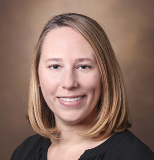
Junzhong Xu, PhD, Director of Cancer Imaging Research at the Vanderbilt University Institute of Imaging Science (VUIIS), is principal investigator of the project. He will work with VUIIS Director John Gore, PhD, and with grant co-investigator Ryan Robison, PhD, MR Clinical Science Leader for North America at Philips Healthcare.

The investigators will integrate the technique into existing clinical MRI systems and develop a quality control system to support consistent use across scanners and institutions. They will then work with researchers from the University of Washington in Seattle to evaluate this technology in clinical breast cancer imaging.

This project “represents an important step in translating a Vanderbilt-developed imaging technology into a tool that could eventually be used in routine clinical care,” Xu said. “Working closely with Philips allows us to address the technical challenges required to move the method beyond a research environment and toward broader clinical use.”

Despite significant advances in early detection and treatment in recent years, breast cancer remains a leading cause of cancer mortality in women. Tissue biopsies provide essential diagnostic information but may provide an incomplete picture of the tumor and are difficult to repeat frequently over the course of treatment.

MR cytometry uses advanced diffusion MRI technology, including oscillating gradient spin echo, to probe how water molecules move within tumors at micrometer length scales. Advanced data acquisition and analysis of this movement enable researchers to estimate mean cell size and cell density noninvasively.

Combining biopsy findings with MR cytometry could give clinicians a more complete picture of a tumor and potentially help guide treatment decisions.

“The ability to measure cell size, cell density and other microstructural features using oscillating gradient diffusion imaging is immensely exciting, given its potential to monitor disease progression and inform clinical decision-making,” Robison said.

“Working alongside Dr. Xu and the team at VUIIS is a tremendous privilege, as they are the pioneers of this technique and are widely recognized as the world leaders in its development, validation and clinical translation,” he said.

“We have had a strong relationship with Philips for over 20 years, and it is encouraging to obtain funding that will be of mutual benefit and help patients,” said Gore, University Distinguished Professor of Radiology and Radiological Sciences, and holder of the Hertha Ramsey Cress Chair in Medicine.

Xu, Associate Professor of Radiology and Radiological Sciences, said MR cytometry has the potential to be applied to imaging other cancers and diseases in which changes in cellular microstructure are important.

The NCI Academic-Industrial Partnership grant (R01CA308592) supporting the research is designed to move promising technologies toward clinical use through collaborations between academic and industry partners.

The post Vanderbilt Health receives $3.4 million grant to advance breast cancer imaging appeared first on Vanderbilt Health News.

Lucy Spalluto and Jennifer Lewis awarded $1 million lung cancer screening grant

Submitted by vicc_news on

Vanderbilt Health’s Lucy Spalluto, MD, MPH, professor of Radiology and Radiological Sciences, and Jennifer Lewis, MD, assistant professor of Medicine in the Division of Hematology and Oncology, have received a grant from AstraZeneca to understand and improve veteran access to mobile lung cancer screening.

Lucy Spalluto, MD, MPH

The study, “REACHing veterans at high risk for lung cancer outside the guidelines and through mobile screening,” will receive approximately $1 million in total grant funding over a four-year period. As explained by Spalluto and Lewis, lung cancer is the leading cause of cancer death in the United States, with incidence higher in veterans compared to the civilian population.

“Improving access to lung cancer screening for a broader population, including those who live in rural areas, through mobile services can increase the early detection of lung cancer and improve lung cancer outcomes,” Spalluto says. “The REACH study explores the impact of mobile lung cancer screening in the Veterans Health Administration. An important component of the REACH study is understanding veterans’ perspectives of mobile screening.”

Jennifer Lewis, MD
Jennifer Lewis, MD

Screening for lung cancer with low-dose CT scans is an effective strategy to detect lung cancer early and improve mortality. However, this screening is widely underutilized, including in the Veterans Health Administration. Veterans living in rural areas are less likely to be screened for lung cancer, and individuals living in rural areas have higher mortality from lung cancer compared to those who live in nonrural areas.

In response to the low screening and high mortality rates, the VA’s Midsouth Veterans Integrated Service Network has partnered with the VA Lung Precision Oncology Program to offer mobile lung cancer screening to better reach veterans who reside in rural areas. Spalluto and Lewis have been awarded the REACH grant through AstraZeneca to understand the reach of mobile lung cancer screening among veterans within and outside current screening eligibility criteria, as well as veterans’ experiences with mobile lung cancer screening.

This work is particularly pertinent to veterans who may have been exposed to war-related chemical, waste and other similar smoke inhalation.

“Exposures that veterans have had because of their military service, such as Agent Orange and burn pits, may place them at high risk for lung cancer,” Lewis explains. “This study will help us understand how many veterans are eligible for lung cancer screening not only based on age and smoking history, but also other important risk factors, such as family history, personal history of cancer, diagnosis of COPD and military environmental exposures. These data will be critical for VA leadership.”

The post Lucy Spalluto and Jennifer Lewis awarded $1 million lung cancer screening grant appeared first on VUMC News.

Women who receive mammograms also undergo lung scans if notified of eligibility

Submitted by vicc_news on

While most women over age 50 schedule mammograms for breast cancer, only a minority who are also eligible for low-dose CT scans for lung cancer undergo those potentially lifesaving screenings. A new study shows that targeted outreach can close the gap.

The study results, published Dec. 1 in the Journal of the American College of Radiology, showed that the improvement in lung cancer screenings exceeded the target enrollment set by the researchers. The study also demonstrated that two different types of outreach initiatives were effective in increasing uptake. Called CALM, an acronym for Coordinate A Lung screening with Mammography, the study was funded by the American Cancer Society.

At one academic medical center, a research team from pulmonary medicine determined eligibility by manual review of smoking history in electronic health records and then contacted patients directly to inform them of eligibility. The team also conducted surveys at mammography locations about smoking history. They had a target enrollment of 200 new patients for lung cancer screening and exceeded it by enrolling 214 patients.

At the other academic center, researchers from radiology identified patients eligible for lung screenings one month prior to their mammography appointments through a review of electronic health records. The patients were offered the opportunity to have both cancer screenings on the same day at the same location. They also exceeded their target enrollment of 322 patients by enrolling 445 patients.

“For years, we have recognized that many women screened for breast cancer are in fact dying from lung cancer. This study allowed us the opportunity to inform women and their referring providers of lung screening eligibility and to facilitate lung screening exams.  We are incredibly grateful for the American Cancer Society and for the National Lung Cancer Roundtable as they supported this multicenter initiative.  We plan to continue these efforts at VUMC and with institutions across the country to save more lives,” said Kim Sandler, MD, professor of Radiology and Radiological Sciences at Vanderbilt University Medical Center, director of the Vanderbilt Lung Screening Program, and the study’s corresponding author and co-principal investigator.

A previous study revealed that 58% of women who were eligible for lung cancer screening had reported having a mammogram within two years compared to only 7.9% who underwent lung cancer screening. Overall, participation in lung cancer screening by both men and women is low, with less than 20% of those eligible for low-dose CT scans receiving the screenings.

“It has been more than 10 years since annual screening for lung cancer was recommended, and screening rates still are disappointingly low. There are many reasons for these low rates, but mostly identifying eligible individuals is challenging in the primary care setting, and there is evidence showing a surprising lack of awareness about lung cancer screening among eligible individuals. The CALM model demonstrates we can successfully recruit eligible women through mammography screening. Perhaps we also will be able to enlist them to encourage eligible family members to have a conversation about lung cancer screening with their health care providers. There is enormous potential here, and the ACS is thrilled with the outcome of this study,” said Robert Smith, PhD, senior vice president and director of the American Cancer Society Center for Early Cancer Detection and the study’s other co-principal investigator.

The researchers hypothesized that mammography screening could be a “teachable moment” for women who are also eligible for lung cancer screening. The study period was from November 2019 to December 2021, but data from 2020 was excluded because of the disruptions in health care screenings due to the onset of the COVID-19 pandemic. Initially, women were considered eligible for lung cancer screening from ages 55 to 80 with a 30 pack-year history of smoking; guidelines expanded in 2021, and women were eligible beginning at age 50 with a 20 pack-year history. One pack year is equal to smoking an average of 20 cigarettes, or one pack, every day for a year. A person who has smoked half a pack per day for 30 years has a 15 pack-year history.

Other VUMC authors on the study are Caroline Godfrey, MD, MPH, Valerie Welty, PhD, Stephen Deppen, PhD, MA, Alexis Paulson, MS, Shanna Joyner, Hannah Marmor, MD, MPH, Grace Wallace, CCRC, Lauren Hatcher, MD, MBA, Landon Fike, MD, and Arulita Gupta, MD.

The post Women who receive mammograms also undergo lung scans if notified of eligibility appeared first on VUMC News.

Subscribe to Department_of_Radiology_and_Radiological_Sciences