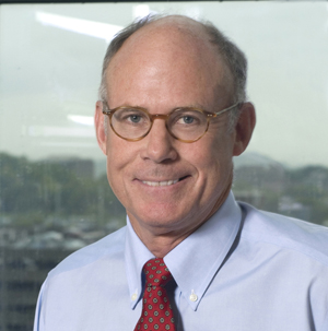
Bhagirathbhai Dholaria, MBBS, Associate Professor of Medicine and a hematopoietic cell transplantation specialist, along with peers from several other transplant centers across the country, recently reached a major milestone in stem cell transplantation.
Bhagirathbhai Dholaria, MBBS
Their clinical trial, called ACCESS, showed that using “mismatched donors” for blood or bone marrow transplants is safe, potentially opening the door to cures for blood cancers such as leukemia and lymphoma.
Findings from the ACCESS trial were recently published in the journal JAMA Network Open.
“Many patients need a donor whose tissue type closely matches their own,” Dholaria said. “Historically, patients from racial and ethnic minority groups have had a harder time finding a perfectly matched donor because donor registries included fewer people from their backgrounds.
“As a result, minority patients are referred less often or move more slowly toward transplant because finding a fully matched donor can be difficult. If doctors believe a suitable donor is unlikely, they may never see a transplant physician in the first place.”
Expanding access and opportunity at the same time
The ACCESS trial set out to dismantle this barrier by allowing mismatched, unrelated donors for patients lacking a full match. Typically, traditional clinical trials restrict enrollment to matched donors only.
By eliminating the requirement for a perfectly matched donor, ACCESS opened the door for more Asian, Black, Hispanic and multiracial patients who previously may not have been eligible for a transplant or clinical trial.
Nearly half of participants in this trial identified as members of racially or ethnically underrepresented groups.
Comparing nearly 670 patients across three different studies, Dholaria and colleagues found that ACCESS participants were more than 25 percentage points more likely to come from diverse racial and ethnic backgrounds than those enrolled in other trials. The broader eligibility criteria also expanded the pool of potential donors available to patients.
“The more we broaden access to transplant, the more patients we can give an opportunity for cure,” said Dholaria.
The University of California, Santa Cruz Alumni Association has announced Melinda Aldrich, PhD, MPH, Professor of Medicine in the Division of Genetic Medicine and Clinical Pharmacology at Vanderbilt Health, as one of its 2026 UC Santa Cruz Alumni Awards recipients.
The awards celebrate individuals whose vision, leadership and dedication have made a meaningful impact in their professions, in society, within the university community, and across the globe. Each honoree exemplifies the values and spirit of UC Santa Cruz through their achievements and contributions, according to a release from UC Santa Cruz.
Aldrich, who has authored more than 170 scientific publications and held leadership roles in numerous national and international scientific organizations, joins John Torigoe, James Hein, PhD, Peter Coha, Jim Gunderson, JD, and Stephanie Barron Lu as recipients of the 2026 UC Santa Cruz Alumni Awards; they will be honored Oct. 16 at a UC Santa Cruz celebration gala.
“This year’s Alumni Award honorees reflect the remarkable breadth of what UC Santa Cruz alumni go on to do and the many ways they contribute to their communities and the world,” said Chancellor Cynthia Larive, PhD. “Their accomplishments are distinct, but each has brought curiosity, purpose, and a commitment to making a difference to their work. We are proud to recognize them and celebrate the paths they have taken since their time at UC Santa Cruz.”
Aldrich, who is also Professor of Biomedical Informatics and Thoracic Surgery at Vanderbilt Health, received her BA in biology from UC Santa Cruz in 1992. She is an internationally recognized expert in lung cancer epidemiology with a research focus on lung cancer screening, risk prediction for early detection, genetics, and differences in health outcomes.
She contributed to health policy revisions of national lung cancer screening guidelines by demonstrating that high-risk African American individuals were disproportionately missed by existing screening eligibility criteria, earning her the Vanderbilt Chancellor’s Award for Research.
“I am deeply honored to receive the UC Santa Cruz Ethos Award. UC Santa Cruz helped shape my belief that scientific progress should benefit all communities, and that principle has guided my work,” said Aldrich, who holds the Robert A. Goodwin Jr., MD, Directorship at Vanderbilt Health. “To be recognized for that commitment by UC Santa Cruz, an institution that has meant so much to me, is especially meaningful.”
Researchers at Vanderbilt Health have found that a balance of two closely related cell surface proteins may be critical to the health of the epithelial lining of the gastrointestinal tract, and for suppressing tumor formation in the colon.
The surface proteins are LRIG1, a tumor suppressor, and its cousin LRIG3, for which there have been conflicting reports about whether it cooperates with or opposes the action of LRIG1.
“We show that a balancing act between LRIG1 and LRIG3 keeps a powerful driver of colorectal cancer in check,” said Robert Coffey Jr., MD, Ingram Professor of Cancer Research and Professor of Medicine. “This offers potentially new avenues to explore in the treatment of colorectal cancer.”
Robert Coffey Jr., MD
Coffey is corresponding author of the report published Aug. 31 in the journal Cellular and Molecular Gastroenterology and Hepatology. Co-director of the Vanderbilt Epithelial Biology Center, he is internationally known for his research on mechanisms that drive the development of colorectal cancer, one of the nation’s leading cancer killers.
In 2012, Coffey and his colleagues found that removal of one copy of the Apc gene, the colon cancer “gatekeeper,” in LRIG1-expressing cells results in multiple colonic tumors.
Now they show that removal of both copies of theLrig3 gene in this setting drastically reduces tumor formation, highlighting the antagonistic nature of the LRIG proteins. This suggests that LRIG1 and LRIG3 work together to maintain the balance of cell signaling pathways implicated in cancer.
Matthew Bechard, PhD, was the paper’s first author. Other Vanderbilt co-authors were Zhengyi Chen, Ping Zhao, PhD, Radhika Aramandla, Yilin Yang, Alan Simmons, Kay Washington, MD. PhD, Jeffery Franklin, PhD, and Ken Lau, PhD.
The research was supported in part by the National Cancer Institute (grant R35CA197570), a GI SPORE grant (P50CA236733), and by the Robert J. Kleberg, Jr. and Helen C. Kleberg Foundation.
For years, care incidence gaps were chalked up to money, insurance and getting to the doctor. While those factors are important, they miss one critical detail: that genes and ancestry shape how cancer develops, proving that no two patients are exactly alike.
“Often, we assume that the reason for most gaps in health care outcomes is an inability to access health care, as some patients simply cannot get transportation to get to the hospital or doctors’ appointments, or they lack insurance,” said Somedeb Ball, MBBS, Assistant Professor of Medicine in Hematology/Oncology at Vanderbilt-Ingram Cancer Center. “But in addition to those factors, we must recognize scientifically that we are not all the same. Understanding the biologic differences in genes and ancestry can provide insights into delivering personalized care.”
Ball, who studies genetic mutations in acute myeloid leukemia (AML), an aggressive blood cancer that has a five-year survival rate of only 30%, set out to investigate and get some answers.
The Problem: A Long-Standing Blind Spot
AML is predominantly diagnosed in white populations, and in turn, up to 90% of clinical trial participants are white. As a result, standard classification criteria, risk stratification models, and approach to targeted therapies have been heavily skewed toward European ancestry, which left big gaps in care for patients of color, particularly Black patients.
But AML is driven by complex combinations of multiple genetic mutations rather than a single defect, so targeted therapies designed around European genetic profiles often fail or lead to treatment resistance in other races and ethnicities.
To address this problem, Ball collaborated with Chidiebube Ugwu, MD, of Jefferson Einstein Philadelphia Hospital, and Jude Ossai, MD, of Newark Beth Israel Medical Center. The research team analyzed American Association for Cancer Research (AACR) Project Genomics Evidence Neoplasia Information Exchange (GENIE) data from 2,359 patients with AML (243 Black and 2,116 white) to examine how mutational landscapes differ by race.
The Key Findings: A Distinct Biological Profile
The study revealed significant biological differences in Black AML patients that directly challenge current “race-blind” risk models:
Lower splicing factor mutations: Black patients exhibited lower rates of splicing factor gene mutations compared to white patients.
Unexpected early-onset mutations: Black patients under age 40 showed a significantly higher prevalence of myelodysplastic syndrome-related mutations, such as ASXL1.
Unique co-mutations: Distinct co-mutation patterns were identified in Black patients, particularly involving the RAS/MAPK signaling pathway.
Altered prognostic indicators: Mutations traditionally classified as “favorable” (such as NPM1) provided protective survival benefits in white patients but failed to do the same in Black patients.
“Because existing risk stratification models were built on data from white patients, we must update how we categorize risk to ensure precision care for Black patients,” Ball said.
The Solutions: Re-Evaluating Guidelines and Expanding Clinical Access
These findings underscore that AML cannot be treated with a “one-size-fits-all” strategy. Shared at the 2026 ASCO Annual Meeting and featured in Specialty Pharmacy Continuum, the team’s research highlights an urgent need for the academic community to incorporate ancestry-specific differences into clinical guidelines.
Moving forward, Ball cited two changes that would work toward a more full understanding of AML: actively recruiting a broad and representative cohort of participants in clinical trials, while expanding genomic testing in community settings.
“Expanding access to genomic tools helps doctors understand the drivers of each patient’s cancer, while robust trial participation ensures new therapies benefit everyone and are applicable to people of all racial and ethnic groups,” he said.
Achieving this will require proactive outreach.
“It is our responsibility as investigators and physicians to meet patients where they are, address their concerns, and clearly communicate how participating in research helps both them and future patients. Building truly inclusive precision oncology requires a unified, comprehensive effort from the top down,” Ball said.
Vanderbilt-Ingram Cancer Center has spent years assembling cancer teams — medical oncologists, surgeons, radiologists, pathologists — in Tullahoma, Spring Hill, Wilson County and beyond.
Proximity is a priority. Advances in immunotherapy and targeted treatment mean many cancers, even advanced ones, are now managed for years rather than months, meaning patients can spend a lot of time on the road to see their care team.
“The goal of treatment is to facilitate your life, not to put your life on hold,” said Laura Goff, MD, MSCI, Executive Medical Director of the Cancer Patient Care Center.
Goff and Waleed Khalaf, MD, PhD, Medical Director for Regional Network Cancer Care, talk about why the evolution of cancer treatments makes mid-state locations essential.
Next month, Community Impact will introduce the people making that shift possible — some of whom grew up in the towns they now practice in. We’ll spotlight the team behind Vanderbilt-Ingram’s regional cancer clinics and revisit the milestones that got us here, such as Cool Springs, which opened in 2001; Wilson County’s radiation oncology growth; and Clarksville’s decades-old radiation program. … And more.
Comprehensive cancer care is available at Vanderbilt Health Pleasant View is located just off Interstate 24 at 6536 Highway 41A in Pleasant View, Tennessee. (photo by Donn Jones)
Q: If you were talking at a Chamber of Commerce in Middle Tennessee about the latest generation of cancer care and how Vanderbilt-Ingram Cancer Center impacts their community, what would you say?
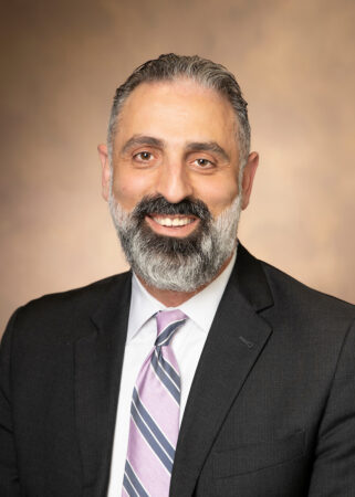
Waleed Khalaf
Goff: Cancer care is impacting everybody’s community. At this point, Vanderbilt-Ingram sees patients who come from great distances to access leading-edge cancer therapies. Cancer care delivery requires a mix of groundbreaking technology and attentiveness, providing much of that care within the context of a patient’s everyday life. Their life goes on. They need to try to work. They need to try to take care of their kids. We want that for them.
Often cancer care goes on for weeks and weeks, and many times even years, so traveling three, four hours round trip on a weekly basis can be a strain on patients. What we are trying to do, Waleed and I together, is get the services we can as local as we can to facilitate the highest quality care in Tennessee towns.
The highly specialized treatments will be done at the Main Campus in Nashville . Thankfully, those are not usually treatments that are needed continually long term or able to be offered everywhere. The value of having Vanderbilt-Ingram as your local provider is the seamless transition to those highly subspecialized things that exist within our network. If you go to Vanderbilt-Ingram, we have that deep expertise. We don’t have to refer you somewhere else.
Khalaf: The main point of emphasis that we would want to highlight is exactly that — bringing that care closer to where our patients live outside of the Main Campus. If you’re somebody living in Spring Hill, for example, we’re in your backyard, and we’re bringing that care closer to you. Cancer care nowadays is provided by a team, of course. It’s not just one person, so we bring the whole team to our regional clinics.
There’s this idea floating around that specialists are only available in Nashville. But we want to underline that Vanderbilt-Ingram specialists are around Middle Tennessee. Your care journey can start with us — and should start — with our specialists who are down the street.
Officials cut the ribbon to mark the opening of the new Vanderbilt-Ingram Cancer Center Lebanon. (photo by Donn Jones)
Q: What does it mean to bring that kind of hope to people who perhaps didn’t think this kind of care was available in the neighboring county?
Goff: Waleed does it and sees it every day. Our new acquisition in Clarksville, the excitement in patients I have seen on campus was palpable as soon as that was announced. They were so excited that Vanderbilt Health was coming to their community. We have a history of people driving from Clarksville, from Owensboro, and beyond. So, to know that Vanderbilt Health was investing in turning around a hospital — we certainly did it in Wilson County and the other locations too, and they were thrilled.
Khalaf: I think that’s exactly right. There are always sensitivities, I think, when we bring a big name to a small community. There are always challenges navigating that. The people, yes, they are excited, very open, but there are some fixtures in the community that are always sensitive and think you’re coming to just bulldoze and take over and change everything and fire everybody here. And that’s not the case. We’ve proven that in Wilson County especially, which has been a tremendous success.
Vanderbilt-Ingram Cancer Center at Spring Hill (photo by Anne Rayner)
Q: How has cancer care evolved in the last, say, 10 to 15 years — and is that partly a reason why decentralizing the care is possible now?
Khalaf: The model of driving three hours to a big hospital to get care is changing. People expect to get care closer to where they live, and we’re trying to match that expectation. At the same time, things are changing dramatically at the level of therapeutics and advances. People are living longer and longer with cancer, and it’s really becoming a chronic medical problem as treatments evolve.
A lot of our treatments are now non-chemotherapy treatments; they’re targeted treatments. A lot of them are pills people take at home — they don’t have to come in for an infusion. A lot of them are immunotherapy treatments. A lot of them enable our patients to continue to live life as close to normal as possible while they are dealing with this chronic illness and receiving treatment.
Goff: What we’re really trying to look at is how we can fit more seamlessly into a patient’s life. I tell patients that the goal of treatment is to facilitate your life, not to put your life on hold. If you have a cancer that is very treatable but not curable, you’re going to be on treatment for a long time. We have to find a way to make treatment fit into your life, without placing any more burden on you than is already there.
This a team sport — it’s not just one oncologist who can alone provide adequate cancer care. The Vanderbilt-Ingram model is that we rely on a team of doctors, our surgical oncologists, our expert radiologists, the deep pathology expertise.All of those specialties are necessary to really take adequate care of a cancer patient. It’s one of the values of getting your care at a Vanderbilt Health system rather than a one-off doctor’s office.
Vanderbilt Health has received a five-year, $3.4 million Academic-Industrial Partnership grant from the National Cancer Institute to support development of magnetic resonance (MR) cytometry as a new tool for breast cancer imaging.
MR cytometry is a magnetic resonance imaging technology pioneered by Vanderbilt Health investigators that provides information about tumor microstructure which cannot be obtained from conventional MRI.
Junzhong Xu, PhD, Director of Cancer Imaging Research at the Vanderbilt University Institute of Imaging Science (VUIIS), is principal investigator of the project. He will work with VUIIS Director John Gore, PhD, and with grant co-investigator Ryan Robison, PhD, MR Clinical Science Leader for North America at Philips Healthcare.
The investigators will integrate the technique into existing clinical MRI systems and develop a quality control system to support consistent use across scanners and institutions. They will then work with researchers from the University of Washington in Seattle to evaluate this technology in clinical breast cancer imaging.
This project “represents an important step in translating a Vanderbilt-developed imaging technology into a tool that could eventually be used in routine clinical care,” Xu said. “Working closely with Philips allows us to address the technical challenges required to move the method beyond a research environment and toward broader clinical use.”
Despite significant advances in early detection and treatment in recent years, breast cancer remains a leading cause of cancer mortality in women. Tissue biopsies provide essential diagnostic information but may provide an incomplete picture of the tumor and are difficult to repeat frequently over the course of treatment.
MR cytometry uses advanced diffusion MRI technology, including oscillating gradient spin echo, to probe how water molecules move within tumors at micrometer length scales. Advanced data acquisition and analysis of this movement enable researchers to estimate mean cell size and cell density noninvasively.
Combining biopsy findings with MR cytometry could give clinicians a more complete picture of a tumor and potentially help guide treatment decisions.
“The ability to measure cell size, cell density and other microstructural features using oscillating gradient diffusion imaging is immensely exciting, given its potential to monitor disease progression and inform clinical decision-making,” Robison said.
“Working alongside Dr. Xu and the team at VUIIS is a tremendous privilege, as they are the pioneers of this technique and are widely recognized as the world leaders in its development, validation and clinical translation,” he said.
“We have had a strong relationship with Philips for over 20 years, and it is encouraging to obtain funding that will be of mutual benefit and help patients,” said Gore, University Distinguished Professor of Radiology and Radiological Sciences, and holder of the Hertha Ramsey Cress Chair in Medicine.
Xu, Associate Professor of Radiology and Radiological Sciences, said MR cytometry has the potential to be applied to imaging other cancers and diseases in which changes in cellular microstructure are important.
The NCI Academic-Industrial Partnership grant (R01CA308592) supporting the research is designed to move promising technologies toward clinical use through collaborations between academic and industry partners.
The Neurofibromatosis and Schwannomatosis Patient and Family Symposium will be held virtually Saturday, Aug. 22, from 9 a.m. to noon. The symposium is free and open to people affected by neurofibromatosis and schwannomatosis, as well as their families, loved ones and caregivers.
Neurofibromatosis (NF) and schwannomatosis are genetic disorders that predispose patients to develop tumors of the nervous system. NF1, the most common form of neurofibromatosis, affects approximately 1 in 2,500 births. Tumors associated with these conditions can occur throughout the nervous system, including the brain, spinal cord and peripheral nerves. Most are benign, although a small number can become cancerous.
Alex Mohler, MD, MS, Associate Professor of Clinical Neurology in the Division of Neuro-Oncology and Director of the Vanderbilt-Ingram Cancer Center Neurofibromatosis Clinic is the symposium chair.
“Until recently, we have had very few options for managing the challenges associated with these conditions,” Mohler said. “Our annual symposium brings the latest information directly to patients and families in our region who are affected by NF and schwannomatosis.”
Participants will receive updates on current treatment options, clinical programs and research initiatives available through Vanderbilt-Ingram. The event will highlight support services available for adults and children living with NF and schwannomatosis. Attendees will also learn about resources available through Vanderbilt Health partners NF Tennessee and the Children’s Tumor Foundation.
Jason Colee, a husband and father of two from Alabama, runs because it refreshes him physically and mentally. In the last three years alone, he has run (and won) multiple 5Ks, several 10Ks, three half-marathons, a 16.2-mile challenge in the Great Smoky Mountains, and even a full marathon.
Notably, just before all these achievements, he underwent a six-hour spine surgery at Vanderbilt University Hospital to remove a rare tumorous mass that was nestled inside his spinal cord.
Colee first noticed problems with his gait, was seen by his orthopedist and tried physical therapy. But after being diagnosed with a myxopapillary ependymoma, Colee’s doctors in Huntsville told him he needed to be seen by the best spine specialists there were.
“Fortunately, we live in North Alabama, and we know Vanderbilt’s got a stellar reputation,” said Colee.
He asked his doctor how soon he should be seen at Vanderbilt Health. “You need to be in there immediately,” was the doctor’s reply.
High risk, high reward
The following week, Colee and his wife, Jennifer, traveled to Vanderbilt-Ingram Cancer Center Belle Meade. He was quickly matched with Scott Zuckerman, MD, MPH, who Colee’s referring physician said would be the best neurosurgeon to handle his case.
“We took it day by day,” said Zuckerman. “We told him it’s a big, high-risk surgery. I couldn’t make any promises about what type of tumor it was or how tightly it was stuck to his spinal cord, but I think he had come to terms with that.”
Colee was admittedly very nervous. But in undergoing the surgery, he gave himself the best shot to preserve his ability to walk, which mattered for two reasons: his love of running and his family.
“There’s a chance that, at some point, you’re going to walk your daughter down the aisle and be there for your grandkids someday, hopefully,” Colee recalled his wife saying. “We’ve got to do this.”
Colee said the “we” mentality Jennifer emphasized helped restore his confidence that the surgery was the right course of action. He credited Jennifer’s resilience and the shared Christian faith of the family — which includes daughter, Emma, and son, Jake — for his ability to withstand the emotional impact of the ordeal.
“The risks were high, but the rewards were high also,” Colee said.
Jason Colee celebrates after completing the Race to Space Marathon, a milestone in the active life he worked hard to regain after wondering whether he would ever walk again.
Nervous about his nervous system, Colee found his care team rose to the occasion
It makes sense that someone headed to anesthesia for spine surgery would be uneasy. Colee’s worry remained despite what he called excellent preoperative care. That’s when Zuckerman came to speak to him.
“This is the A-team,” Zuckerman told Colee. “Everybody you see in this room is the best at what they do.”
On one hand, getting the best of the best may signal that this is as serious as it gets. But on the other, it’s the ultimate assurance that a patient has found the right surgical team.
“The operating room is a serious, focused environment,” Zuckerman said. “But we go into any complex surgery with a really good plan, just like a coach would go into a basketball game with a focused game plan.”
Colee said he never fully conquered his nerves before the surgery.
“Vanderbilt is first-rate,” Colee said. “The process of getting there and everything they did to get me back and ready to go was top-notch; unlike anything we’d ever experienced. It was fantastic.”
Finally, thanks to the anesthesia, his worries subsided as he slipped into sleep. When he awoke, with the tumor out of his spine, he was hit with a wave of relief. Motor function testing showed that everything came back clear.
“I shouldn’t be this excited about being able to wiggle my dang toes,” Colee said. “I guess the gravity of it all hit me after the fact.”
Jason Colee walks his daughter, Emma, down the aisle on her wedding day. Moments like this one were top of mind for Jason and his wife, Jennifer, as they weighed the risks of spinal surgery.
Life after a spinal tumor was just as fulfilling as before
Colee was grateful that his rehabilitation care team wanted input on how he wanted to attack recovery.
“‘We’ve got guidelines we want you to follow and milestones we want you to hit, and it’s up to you how fast you hit those,’” Colee recalled being told. “The nurses let me do what I wanted to do when I wanted to do it as long as it was safe.”
In the end, Colee left the hospital only 48 hours after the conclusion of surgery, a remarkable outcome for a spinal operation.
Soon, Colee wondered when he could get back to running. Zuckerman said if he followed his recovery plan to the letter, it wouldn’t be long.
All told, it ended up only being a six-week hiatus before he put his running shoes on again. It was ugly, Colee said, “like a frog in a frying pan,” but he was moving.
Then came the 5Ks, 10Ks, half-marathons, 16.2-mile race and a full marathon this past spring.
But the most important thing Colee did with his legs didn’t come on the racecourse. It was, as his wife had foreseen, walking his daughter down the aisle.
“Everybody at Vanderbilt was invested in us getting back to where we wanted to be,” Colee said. “This was not guaranteed, even with the surgery. But I knew that Dr. Zuckerman and everybody involved in the process wanted to help me do what I wanted to, whether it was walking my daughter down the aisle or checking a marathon off the bucket list.”
Nearly two decades ago, after being diagnosed with leukemia at age 7, Lily Hensiek promised to help other children diagnosed with cancer.
At the time, doctors at Monroe Carell Jr. Children’s Hospital at Vanderbilt explained her diagnosis through a simple analogy. Cancer was like weeds taking over a garden, they told her, and chemotherapy was the tool used to remove them. The idea inspired her family to create the Lily’s Garden Fund, a fundraising effort dedicated to advancing childhood cancer research, training and innovation.
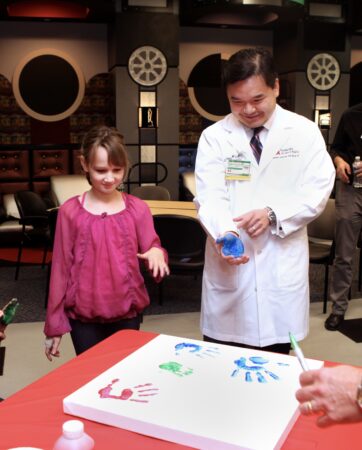
Lily Hensiek serves as patient ambassador for Hyundai Hope on Wheels event at Monroe Carell in 2011. (photo by Susan Urmy)
Over the past 18 years, Lily’s Garden has funded pediatric cancer research, programs, lectureships, fellowship training and early-career investigators.
The family recently announced its latest commitment: $5 million to establish the Lily’s Garden Pediatric Leukemia Program at Monroe Carell to support leukemia research, physician-scientist leadership, precision oncology, innovative discovery projects and family-centered care initiatives. This gift brings the family’s total giving to $8 million. Family members have added to Lily’s Garden through personal giving, fundraising events, and the support of friends and community partners.
“We are so incredibly grateful to Lily’s family; they are absolutely amazing,” said Debra Friedman, MD, MS, Director of the Division of Pediatric Hematology and Oncology at Monroe Carell and Deputy Director of Vanderbilt-Ingram Cancer Center.
“This leukemia program will provide us the ability to continue to advance discovery for leukemia and then translate that discovery to patients in the clinic,” continued Friedman, holder of the E. Bronson Ingram Chair in Pediatric Oncology.
A Family Affair
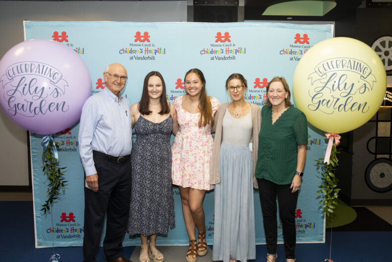
The Lily’s Garden Fund has been a family affair. Joining Lily in the endeavor are her parents, Larisa and Phillip Featherstone; grandparents, Carol and Ron Johnston; and sisters, Sophie Hensiek and Sophia Featherstone.
Lily and Sophie Hensiek make soaps for their first fundraiser for pediatric cancer efforts. (submitted photo)
The family, from Franklin, Tennessee, owns a workplace safety consulting and third-party claims administration firm, Johnston & Associates, which was founded by Ron.
Lily was first diagnosed with pre-B cell acute lymphoblastic leukemia when she was in second grade. At the time, she set what felt like a lofty goal — raising $1 million for childhood cancer research.
“I never imagined when we started the Lily’s Garden Fund that it would go this far or become this big,” said Hensiek, who is now 25. “I’m very thankful that we’ve had the opportunity to do all of this and help support research.”
The family’s philanthropy journey began simply.
“Our first fundraiser was making soap,” said Larisa Featherstone, who also serves as Chair of the Monroe Carell Advisory Board. “Honestly, it felt insurmountable,” Featherstone said. “But it shows how something really small can compound over time, and every bit truly makes a difference.”
Left to right: Ron Johnston, Lily Hensiek, Sophie Hensiek, Sophia Featherstone and Carol Johnston celebrate Lily’s Garden efforts at Monroe Carell in 2023. (photo by Susan Urmy)
A New Era of Discovery
The newest gift is designed to accelerate the rapid progress in research. The gift will support a directorship to help advance pediatric leukemia care throughout the care continuum, including long-term survivorship; it will also create a directorship in cancer predisposition and precision oncology; and it will provide resources for innovative, high-impact research projects.
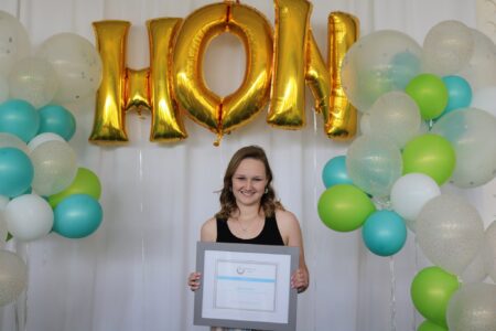
Lily Hensiek was honored for raising awareness and funds for pediatric cancer research at the Mary Catherine Strobel Volunteer Awards in 2019. (submitted photo)
Hensiek knows firsthand how life-changing research can be. As a teen, she experienced a rare late relapse of acute lymphoblastic leukemia and benefited from an immunotherapy treatment previously unavailable when she was first diagnosed.
“Immunotherapy wasn’t really even a thing when I was 7 years old,” Hensiek said. “It’s really amazing to see how far research has moved forward.”
For the type of leukemia Hensiek had, cure rates have risen dramatically.
“When Lily was first diagnosed, we said there was probably somewhere around 85% to 90% cure,” Friedman said. “Now, with the same leukemia, due to advances, we’re at almost 98%.”
The program will also support mental health and family-focused initiatives — areas the family believes are essential.
“Childhood cancer affects the whole family,” Featherstone said. “It doesn’t just affect the child or the parent.”
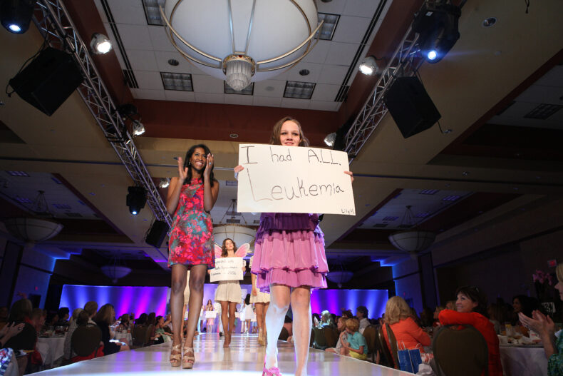
Lily Hensiek models at Friends & Fashion fundraising event in 2013. (photo by Susan Urmy)
A Full-Circle Moment
Today, Hensiek is doing well and preparing for a career in health care. In a full-circle moment, she is studying to be a physician assistant at Kansas State University and will graduate in May 2027. Inspired by the physicians and caregivers who helped save her life, she too hopes to help children with cancer.
She also had the opportunity to do some of her clinical training at Monroe Carell. “It was such a phenomenal experience,” she said. “I’ve been the patient, and now I’ve been the student. That’s kind of wild to think about.”
As Lily’s Garden enters its next chapter, the family hopes the new leukemia program will honor not only Hensiek’s journey, but also the many children and families they have met along the way.
“We just keep going until there’s a cure,” Hensiek said.
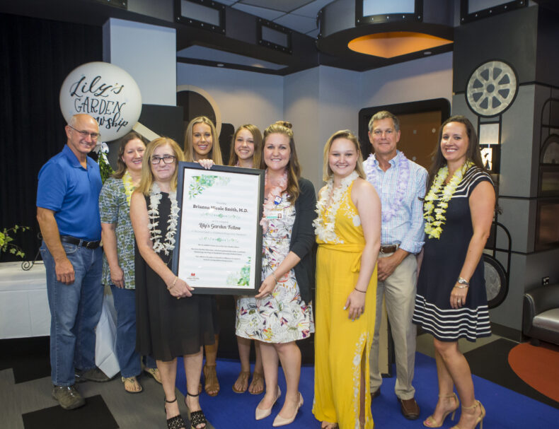
On hand for a celebration of Lily’s Garden initiatives in 2018 were, holding the plaque, Debra Friedman, MD, MS, left, and Brianna Smith, MD, MS; at right, Larisa and Phillip Featherstone with their daughters, Lily, Sophie and Sophia, and at left, Carol and Ron Johnston. (photo by Susan Urmy)
Gastric cancer rarely develops overnight. Instead, chronic inflammation can trigger a series of changes in stomach tissue that progress from normal cells to precancerous lesions and eventually to cancer. But where the mystery lies is why do some precancerous lesions develop into cancer while others do not?
A new study from Eunyoung Choi, PhD, and colleagues helps answer that question by identifying the cancer-associated transcription factor SOX9 as a critical factor in the progression of spasmolytic polypeptide-expressing metaplasia (SPEM), a cell type increasingly recognized as a precursor to gastric cancer. The research was published July 14 in the journal Gastroenterology.
Eunyoung Choi, PhD
“Normal gastric chief cells transition into SPEM cells, which are associated both with mucosal recovery and cancer development,” said Choi, Associate Professor of Surgery and holder of the Naji N. Abumrad Chair. “Although SPEM has been recognized for years, the mechanisms regulating its progression have remained unclear.”
During chronic gastric injury, mature gastric chief cells can transform into SPEM cells as part of the stomach’s repair process. While this response may initially be protective, persistent inflammation and cancer-promoting signals can cause these cells to acquire characteristics associated with cancer development.
To investigate SOX9’s role in this process, researchers used two mouse models of gastric injury and carcinogenesis along with human gastric tissue samples. They found that SOX9 is required for chief cells to transition into SPEM cells following both acute and chronic injury.
When the researchers genetically removed SOX9, metaplasia failed to develop. The loss of SOX9 also prevented progression toward cancer and allowed repair-associated progenitor cells to return to a more normal state.
“These findings suggest that SOX9 functions as a master regulator of the cellular plasticity that drives gastric cancer development,” Choi said.
The study also revealed that SOX9-dependent SPEM cells help recruit fibroblasts to the base of gastric glands, contributing to a tissue environment that supports tumor formation.
Additionally, investigators identified a distinct subgroup of SPEM cells that express TOP2A, a protein associated with cell proliferation. Human tissue analyses showed that cells positive for both SOX9 and TOP2A were linked to metaplastic progression, suggesting these markers may help identify precancerous lesions at greater risk of becoming cancerous.
The findings may have important implications for patient care.
“Finding metaplasia on a biopsy is relatively common, but predicting which lesions will progress to cancer has remained challenging,” Choi said. “SOX9 and TOP2A positivity along with other SPEM cell markers may help identify patients with a higher likelihood of progression.”
Beyond their potential use as biomarkers, the results also point to new therapeutic possibilities. Targeting the cellular pathways regulated by SOX9 could provide an opportunity to interrupt the cancer development process before invasive disease occurs.
The researchers concluded that SOX9 is a critical regulator of SPEM development, metaplastic progression and gastric carcinogenesis. Future studies will explore how these findings can be translated into improved risk assessment tools and preventive strategies for patients at risk for gastric cancer.
The paper’s first author was Alexis Guenther, a graduate student in Cell and Developmental Biology in the Choi lab. Other Vanderbilt co-authors were Amanda Ruelas, Brianna Caldwell, MBA, Youngwon Cho, PhD, Changqing Zhang, Judith Romero-Gallo, MS, and Richard Peek, MD.
The study was supported in part by the National Institutes of Health (grants F31CA284715, R37CA244970, R01CA272687, R01CA077955, P01CA116087, R01DK058587, R01CA281732 and P20GM121322), the American Gastroenterological Association Research Foundation, the U.S. Department of Defense, and the Vanderbilt University School of Medicine Stanley Cohen Innovation Fund.